 Click image to be taken to the source.
 A General Overview
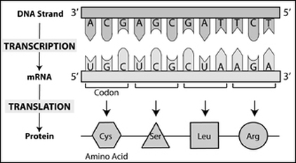
- Translation is the second step of the "central dogma" of biology, following transcription. During this process, ribosomes attach to the mature strand of mRNA produced from DNA transcription to produce a string of amino acids, called a polypeptide chain. This chain is then folded to produce a protein.
 Click image to be taken to the source. Click image to be taken to the source.
|
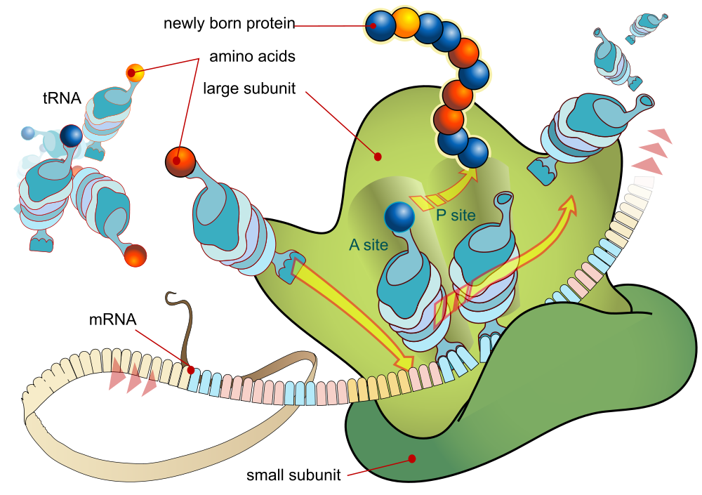
| Messenger RNA (mRNA): mRNA is the single-stranded compliment of the template strand of DNA. The template strand goes from 3' to 5', while the mRNA strand produced goes from 5' to 3'. It caries the protein production information from the nucleus, where transcription takes place, to the cytoplasm, where translation takes place. This strand is composed of nucleic bases adenine (A), guanine (G), cytosine (C), and uracil (U). It is important to note that thymine (T) is no longer present as it was in DNA, instead replaced by uracil (U). A series of three consecutive bases in mRNA is called a codon. There are 64 different codon combinations possible, but these 64 codons only code for 20 amino acids. Of these 64 codons, three are stop codons (UAA, UAG, UGA) and one is the start codon (AUG, methionine). 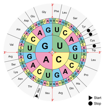 Click image to be taken to the source. A wheel such as the one above is useful to determine which amino acid a codon codes for. | Ribosomal RNA (rRNA): rRNA is the site for protein synthesis and contains the enzymes necessary for the process. The ribosome itself has two subunits, one large and one small, that come together to translate a strand of mRNA. The strand is translated from the 5' end to the 3' end. 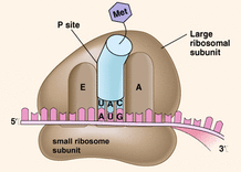 Click image to be taken to the source. Within the ribosome are three sites: the A site, P site, and E site. To remember what occurs at each step, it is helpful to memorize the bolded words in the descriptions below. A site: in the A site, the ribosome ACCEPTS the tRNA molecule with the appropriate anticodon. This anticodon binds to the codon of the mRNA strand. | Transfer RNA (tRNA): tRNA has a set of three nucleotides, called an anticodon, that is complimentary to the codon of the mRNA strand. Attached to the tRNA molecule is the corresponding amino acid. The codon of the mRNA strand and its corresponding tRNA anticodon are bonded with hydrogen bonds following the base pairing principles. The anticodon sits in the anticodon loop, seen at the bottom of the image. The amino acid is attached to the top of the tRNA molecule on the 3' end with an ester bond (-C-O-O-). The amino acids are free-floating within the cytoplasm. 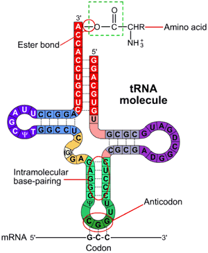 Click image to be taken to the source. The construction of tRNA molecules is done using Aminoacyl-tRNA synthetase, an enzyme that catalyzes the ester bond formation between the tRNA molecule and the amino acid. The resulting complex is called an aminoacyl-tRNA. |
The Steps of Translation
Once the mRNA has been processed in the nucleus, it is able to leave the nucleus to enter the cytoplasm, where tranaslation takes place.
Once the mRNA has been processed in the nucleus, it is able to leave the nucleus to enter the cytoplasm, where tranaslation takes place.
- Initiation: The 5' end of the mRNA with the cap is scanned by the small ribosomal subunit until the start codon, AUG, is recognized. The aminoacyl-tRNA molecule with the amino acid methionine binds to the codon, and the large subunit will attach to the small subunit. The aminoacyl-tRNA with methionine attached to it is in the P site.
- Elongation: The next codon is read and its corresponding aminoacyl-tRNA is bonded to the A site. A peptide bond is formed between methionine and the next amino acid. This entire complex is shifted down so that the tRNA molecule that held methionine is at the E site and the aminoacyl-tRNA molecule with the next amino acid is now at the P site, leaving the A site open for the next aminoacyl-tRNA. The tRNA that held methionine is ejected from the complex to be recycled in the cytoplasm, and the processes repeats until a stop codon is reached.
- Termination: When the ribosome reaches UAA, UAG, or UGA, translation ends. There is no aminoacyl-tRNA molecule that corresponds to the stop codon. Instead, a release factor moves to the A site that signals the release of the polypeptide chain and the dissociation of the small and large ribosomal subunits.
Translation (Elongation)
Click image to be taken to the source.
Click image to be taken to the source.
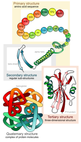 Click image to be taken to the source. | Protein Structures As the polypeptide chain grows, folding is already taking place. This folding action is the distinction between a polypeptide chain and a folded protein. Each distinct fold is significant and precise. The amino acid sequence, without folds, is already considered a primary structure. Once it is folded, it becomes a secondary structure. The polypeptide chain may fold to become either an alpha helix or a pleated sheet. After more folding, it is known as a tertiary structure. If and when two or more polypeptide chains interact with one another in the folding process, the polypeptide chains fold together to become a quarternary structure. |
| The following video is helpful for DNA chromosome wrapping, DNA replication, transcription, and translation. Skip to 4:47 if you wish to only watch the clip on translation. (x) | 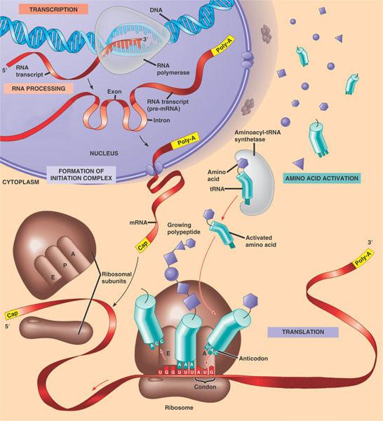 An overview of protein synthesis. Click image to be taken the source. |
